Compact Bone Diagram - 6 3 Bone Structure Anatomy Physiology / 0 0000 a shoutout is a way of letting people know of a.. The periosteum then secretes compact bone superficial to the spongy bone. Label number 1 in the diagram indicates which part of the bone. The new bone is constantly also remodeling under the action of osteoclasts (not shown). Andrew kirmayer a diagram of the anatomy of a bone, showing the compact bone. Compact bone, also called cortical bone, dense bone in which the bony matrix is solidly filled with organic ground substance and inorganic salts, leaving only tiny spaces (lacunae) that contain the osteocytes, or bone cells.compact bone makes up 80 percent of the human skeleton;
Cortical bone is compact bone while cancellous bone is trabecular and spongy bone. It is dense (because of calcified matrix) with tiny spaces known as lucanas. Some, mostly older, compact bone is remodelled to form these haversian systems (or osteons). Microscopic structures of compact bone wedge of bone duration. It is also called osseous tissue or cortical bone and it provides structure and support for an organism as part of its skeleton, in addition to being a location for the storage of minerals like calcium.about 80% of the weight of the human skeleton comes from.
Structure Of Compact Bone Diagram Png Bone Transparent Free Transparent Png Images Pngaaa Com from image.pngaaa.com About press copyright contact us creators advertise developers terms privacy policy & safety how youtube works test new features press copyright contact us creators. A diagram of the anatomy of a bone, showing the compact bone. As seen in the image below, compact bone forms the cortex, or hard outer shell of most bones in the body. The diagram above shows a longitudinal view of an osteon. Compact and spongy.the names imply that the two types differ in density, or how tightly the tissue is packed together. There are small canals that run through the bone, which allow blood vessels to penetrate it. Learn vocabulary, terms, and more with flashcards, games, and other study tools. Andrew kirmayer a diagram of the anatomy of a bone, showing the compact bone.
Connected to surrounding osteoblasts and osteocytes through.
Under periosteum of all bones is the bulk of the diaphysis of long bones. Compact bone diagram osteon compact bone ap pinterest anatomy human anatomy and. Label number 1 in the diagram indicates which part of the bone. Compact bone accounts for 80% of the bones in the human body. There are small canals that run through the bone, which allow blood vessels to penetrate it. The compact bone is composed of calcified extracellular material the bone matrix and 3 major cell types which are osteoblast which ssynthesize and secrete the organic components of bone matrix which include type 1 collagen fibers proteoglycans and several glycoproteins such as ostepnectin. The new bone is constantly also remodeling under the action of osteoclasts (not shown). There are pores and spaces even in compact bone. (b) in this micrograph of the osteon, you can clearly see the concentric lamellae and central canals. Learn vocabulary, terms, and more with flashcards, games, and other study tools. Compact bone is the denser, stronger of the two types of bone tissue ( (figure) ). Cortical bone is compact bone while cancellous bone is trabecular and spongy bone. Although the calls are close together, this type of bone is not completely solid.
It is dense (because of calcified matrix) with tiny spaces known as lucanas. In long bones, as you move from the outer cortical compact bone to the inner medullary cavity, the bone transitions to spongy bone. Compact bone is the denser, stronger of the two types of bone tissue ( (figure) ). Cortical bone is compact bone while cancellous bone is trabecular and spongy bone. Compact bone, also called cortical bone, dense bone in which the bony matrix is solidly filled with organic ground substance and inorganic salts, leaving only tiny spaces (lacunae) that contain the osteocytes, or bone cells.compact bone makes up 80 percent of the human skeleton;
Structure Of Compact Bone Download Scientific Diagram from www.researchgate.net In long bones, as you move from the outer cortical compact bone to the inner medullary cavity, the bone transitions to spongy bone. Long bone labeled compact bone / trabeculae of bone: Compact bone accounts for 80% of the bones in the human body. Diagram of a typical long bone showing both cortical (compact) and cancellous (spongy) bone. A diagram of the anatomy of a bone, showing the compact bone. Compact bone diagram osteon compact bone ap pinterest anatomy human anatomy and. The two main structural components typically include spongy bone on the interior, with an outer layer of compact bone. Andrew kirmayer a diagram of the anatomy of a bone, showing the compact bone.
There are two types of bone tissue:
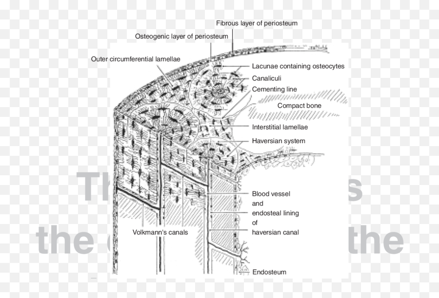
Label femur diagram handout • review the following terms: A diagram of the anatomy of a bone, showing the compact bone. Compact bone is the denser stronger of the two types of bone tissue. It makes up the outer cortex of all bones and is in immediate contact with the periosteum. Compact and spongy.the names imply that the two types differ in density, or how tightly the tissue is packed together. The remainder is cancellous bone, which has a spongelike appearance with numerous large spaces and is found in the. To recognise bone and understand its structure and to understand the processes by which bone can be formed. (b) in this micrograph of the osteon, you can clearly see the concentric lamellae and central canals. Because of its strength, the compact bone makes it possible for the bone to support weight. Connected to surrounding osteoblasts and osteocytes through. There are pores and spaces even in compact bone. The remainder of the bone is formed by cancellous or spongy bone. Compact bone is formed from a number of osteons, which are circular units of bone material and blood vessels.
Andrew kirmayer a diagram of the anatomy of a bone, showing the compact bone. Bone · march 20, 2021. About press copyright contact us creators advertise developers terms privacy policy & safety how youtube works test new features press copyright contact us creators. They allow blood vessels and nerves to travel through them to supply the osteocytes. The remainder is cancellous bone, which has a spongelike appearance with numerous large spaces and is found in the.
Hsc210 Lab Man Lab 3 from www.bio.miami.edu There are pores and spaces even in compact bone. Compact bone, as opposed to spongy bone, is made of cylindrical units, called osteons, that are tightly formed together. Label number 1 in the diagram indicates which part of the bone. Compact bone is formed from a number of osteons, which are circular units of bone material and blood vessels. Unit 3 part 1 x section bone. Compact bone is the strongest form of bone tissue containing few spaces. Related posts of compact bone diagram labeled anatomical diagram of the abdomen. The remainder is cancellous bone, which has a spongelike appearance with numerous large spaces and is found in the.
They allow blood vessels and nerves to travel through them to supply the osteocytes.
Compact bone is the denser stronger of the two types of bone tissue. There are two types of bone tissue: The remainder is cancellous bone, which has a spongelike appearance with numerous large spaces and is found in the. Compact bone diagram bone cross section diagram file624 diagram of compact bone new. 0 0000 a shoutout is a way of letting people know of a. They allow blood vessels and nerves to travel through them to supply the osteocytes. Haversian canals (sometimes canals of havers) are a series of microscopic tubes in the outermost region of bone called cortical bone. About press copyright contact us creators advertise developers terms privacy policy & safety how youtube works test new features press copyright contact us creators. Between the rings of matrix the bone cells osteocytes are located in spaces called lacunae. Anatomical diagram of the abdomen 12 photos of the anatomical diagram of the abdomen anatomical diagram of the abdomen, diagram of the abdomen area, diagram of the abdomen female, diagram of the abdomen muscles, diagram of the abdominal aorta, human anatomy, anatomical diagram of the abdomen, diagram of the. S3.amazonaws.com compact bone consists of outer and inner sheets of lamellar bone (not seen here) and haversian systems, shown here, that run parallel to the long axis of bones. Compact bone is the denser, stronger of the two types of bone tissue ( (figure) ). Compact bone is the denser, stronger of the two types of osseous tissue (figure 6.3.6).